Главная страница Случайная страница
КАТЕГОРИИ:
АвтомобилиАстрономияБиологияГеографияДом и садДругие языкиДругоеИнформатикаИсторияКультураЛитератураЛогикаМатематикаМедицинаМеталлургияМеханикаОбразованиеОхрана трудаПедагогикаПолитикаПравоПсихологияРелигияРиторикаСоциологияСпортСтроительствоТехнологияТуризмФизикаФилософияФинансыХимияЧерчениеЭкологияЭкономикаЭлектроника
Development
|
|
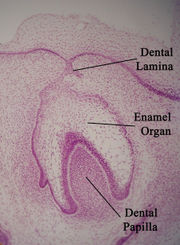

Histologic slide showing a developing tooth. The mouth would be in the area of space at the top of the picture.
Enamel formation is part of the overall process of tooth development. When the tissues of the developing tooth are seen under a microscope, different cellular aggregations can be identified, including structures known as the enamel organ, dental lamina, and dental papilla. The generally recognized stages of tooth development are the bud stage, cap stage, bell stage, and crown, or calcification, stage. Enamel formation is first seen in the crown stage.
Amelogenesis, or enamel formation, occurs after the first establishment of dentin, via cells known as ameloblasts. Human enamel forms at a rate of around 4 μ m per day, beginning at the future location of cusps, around the third or fourth month of pregnancy. As in all human processes, the creation of enamel is complex, but can generally be divided into two stages. The first stage, called the secretory stage, involves proteins and an organic matrix forming a partially mineralized enamel. The second stage, called the maturation stage, completes enamel mineralization.
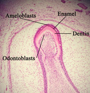
Histologic slide showing enamel formation
In the secretory stage, ameloblasts are polarized columnar cells. In the rough endoplasmic reticulum of these cells, enamel proteins are released into the surrounding area and contribute to what is known as the enamel matrix, which is then partially mineralized by the enzyme alkaline phosphatase. When this first layer is formed, the ameloblasts move away from the dentin, allowing for the development of Tomes’ processes at the apical pole of the cell. Enamel formation continues around the adjoining ameloblasts, resulting in a walled area, or pit, that houses a Tomes’ process, and also around the end of each Tomes’ process, resulting in a deposition of enamel matrix inside of each pit. The matrix within the pit will eventually become an enamel rod, and the walls will eventually become interrod enamel. The only distinguishing factor between the two is the orientation of the calcium phosphate crystals.
In the maturation stage, the ameloblasts transport substances used in the formation of enamel. Histologically, the most notable aspect of this phase is that these cells become striated, or have a ruffled border. These signs demonstrate that the ameloblasts have changed their function from production, as in the secretory stage, to transportation. Proteins used for the final mineralization process compose most of the transported material. The noteworthy proteins involved are amelogenins, ameloblastins, enamelins, and tuftelins. During this process, amelogenins and ameloblastins are removed after use, leaving enamelins and tuftelin in the enamel. By the end of this stage, the enamel has completed its mineralization.
At some point before the tooth erupts into the mouth, but after the maturation stage, the ameloblasts are broken down. Consequently, enamel, unlike many other tissues of the body, has no way to regenerate itself. After destruction of enamel from decay or injury, neither the body nor a dentist can restore the enamel tissue. Enamel can be affected further by non-pathologic processes. The discoloration of teeth over time can result from exposure to substances such as tobacco, coffee, and tea. This is partly due to material building up in the enamel, but is also an effect of the underlying dentin becoming sclerotic. As a result, tooth color gradually darkens with age. Additionally, enamel becomes less permeable to fluids, less soluble to acid, and contains less water.
Dentin
Dentin: (dentine) is a calcified tissue of the body, and along with enamel, cementum, and pulp
is one of the four major components of teeth. Usually, it is covered by enamel on the crown and cementum on the root and surrounds the entire pulp.
By weight, seventy percent of dentin consists of the mineral hydroxylapatite, twenty percent is organic material and ten percent is water. Yellow in appearance, it greatly affects the color of a tooth due to the translucency of enamel. Dentin, which is less mineralized and less brittle than enamel, is necessary for the support of enamel.
Dentin consists of microscopic channels, called dentinal tubules, which radiate outward through the dentin from the pulp to the exterior cementum or enamel border. These tubules contain fluid and cellular structures. As a result, dentin has a degree of permeability which can increase the sensation of pain and the rate of tooth decay..
The formation of dentin, known as etinogenesis, begins prior to the formation of enamel and is initiated by the odontoblasts of the pulp. Unlike enamel, dentin continues to form throughout life and can be initiated in response to stimuli, such as tooth decay or attrition.
There are different types of dentin, differentiated by appearance and stage of development. Primary dentin forms most of the tooth. Secondary dentin develops after root formation is complete and forms much slower than primary dentin. Tertiary dentin forms as a biological response to stimuli.