Главная страница Случайная страница
КАТЕГОРИИ:
АвтомобилиАстрономияБиологияГеографияДом и садДругие языкиДругоеИнформатикаИсторияКультураЛитератураЛогикаМатематикаМедицинаМеталлургияМеханикаОбразованиеОхрана трудаПедагогикаПолитикаПравоПсихологияРелигияРиторикаСоциологияСпортСтроительствоТехнологияТуризмФизикаФилософияФинансыХимияЧерчениеЭкологияЭкономикаЭлектроника
Methods of Examination 4 страница. In liver pathology, the rate and the degree of absorption and discharge of rose bengal decreases
|
|
| fa jo Why 2o\ 7? ifr y\k ' Himi nl in 11 i: 111 i |
| Fig. 96. Accumulation and excretion of rose bengal. a —in norm; b —in hepatitis. |
 In liver pathology, the rate and the degree of absorption and discharge of rose bengal decreases. If polyhedral cells are affected, the absorption process is especially affected. The secretory function of the liver is predominantly affected in inflammation and especially in impaired patency of the bile ducts.

Special Part
Chapter 7. Digestive System

|
|
| I I Iimfi'im ii HI HI 1 HI lit 1111 Illl |
| 11 mtlliPliill II I I III lilBlll I HUM illl I II 1 |
| ____ uwinMWii |
| mmm i m i .JIJUllll II II in mini i r 111! II 111 |
| MSMSSSWf, |

 Fig. 97. Scanogram of a normal liver.
Scanning is a graphic registration of distribution of labelled compounds in the liver (hepatoscanogram). The patient is given intravenously rose bengal labelled with 131I (3 fid/kg) in 0.8-1 ml of isotonic sodium chloride solution (or labelled with 198Au). Scanning is carried out in 30 minutes after the injection.
A scanogram of a healthy subject (Fig. 97) shows distinct borders of the liver and diffuse distribution of the radioactive substance in it. In diffuse affection of the liver (chronic hepatitis, cirrhosis) its contours are irregular and indistinct, the liver shadow is uneven and spotty: areas with normal absorption of the isotope alternate with large areas of decreased radioactivity which indicates dysfunction of the polyhedral cells of the liver (Fig 98). Focal affections of the liver (primary and metastatic cancer, echinococcal cysts) can be seen on a scanogram as defects of absorption of the radioactive substances, i.e. as foci of diffuse thinned shadows. Scanning with radioactive colloidal gold is used to determine total activity of the entire reticulohistiocytic system and the mesenchymal function of the liver and spleen. In healthy people 198Au is accumulated mainly in the liver as compared with the spleen. Absorption of the colloidal gold in an enlarged spleen increases in cirrhosis of the liver (Fig. 98).
Fig. 98. Scanogram in portal liver cirrhosis.
Colour scanning has been used in recent years. Scanograms more vividly show different colouration of various zones due to different accumulation of the isotope in the organs. Scanograms are interpreted quantitatively by special gamma-chambers which record simultaneously radioactivity over the entire organ (without moving the detector over the examined region). The time of examination is thus shortened. Computerized tomography is used in cases where scanning fails to give sufficient information for a correct diagnosis. This, however, is a more complicated method.
ECHOGRAPHY
Echography is widely used in hepatology. Ultrasound can be used to assess the condition of the liver tissue, to detect cysts (almost in 90 per cent of cases), abscesses and tumours of the liver (almost in 80 per cent of cases). Successive use of radioisotope scanning of the liver and echohepatography improves accuracy of diagnosis and facilitates differentiation of focal affections of the liver. Ultrasound helps to perform sighting biopsy of the liver and to differentiate between cirrhosis, hepatitis, and fatty degeneration, and to assess the extent of liver affection. This method can be used to reveal liver affections in a comparatively early stage
Special Part
Chapter 7. Digestive System


 of the process. Ultrasound is used to study the porta hepatis, e.g. to detect dilated and twisted portal vein in portal hypertension. Examination of the spleen establishes its position, reveals possible enlargement (which may be an indirect sign of liver cirrhosis), and determines the structure of this organ.
Echography is especially useful to diagnose diseases of the gall bladder. Position of the gall bladder, the presence of stones in it, and the condition of its walls can be assessed. True, ultrasound is not so effective as cholecystography in revealing cholelithiasis, but it is especially advantageous in cases when cholecystography proves ineffective or intravenous cholecystography becomes impossible due to hyperbilirubinaemia and jaundice which are contraindications to these examinations, or due to allergy to contrast substances, or general grave condition of the patient. Meteorism and much subcutaneous fat (in disorders of fat metabolism) impair accuracy of echographic diagnosis, but these factors interfere also with cholecystographic study of the gall bladder.
A two-dimensional scanning method (B-scanning) can be used to study the common bile duct and sometimes to establish the cause of its obstruction (stones, tumour). Echography is used to diagnose obstruction of the gall bladder by a stone, dropsy or empyema of the gall bladder which arise in such obstructions, and also cancer of the gall bladder, which occurs not infrequently.
PUNCTURE BIOPSY OF THE LIVER
Puncture biopsy is used to take specimens of liver tissue from a patient for histochemical examinations (electron microscopy) and also to study liver enzymes. Puncture biopsy helps in cases where diagnosis of diffuse liver affections is difficult. The procedure is only carried out for special indications. Two methods of biopsy are used: " blind" and sighting (under control of laparoscope). The former method is used in diffuse affections, and the latter in focal affections.e.g. in suspected cancer.
Technique. The patient lies on his back without pillow, slightly turning to his left side with the right arm behind the head. The skin is properly treated and anaesthetized at the site of puncture. A 1 or 2 per cent procaine solution is used for anaesthesia (2-3 ml). Using a stilette, the skin is cut to the depth of 2—4 mm in the region of the 9th interspace, in the anterior axillary line. This is the broadest zone of liver dullness. A special Menghini needle (or its modification according to Bluger and Sinelnikova) with a plunger inside it is introduced into the punctured skin. The needle is connected to a syringe containing a few millilitres of isotonic sodium chloride solution. After puncturing the skin, part of the isotonic solution is
discharged from the syringe in order to expell pieces of skin and subcutaneous fat from the needle. The needle is then introduced by a swift movement perpendicularly to the skin surface into the liver and its tissue is sucked in by the syringe. The obtained sample is quickly placed in a fixing solution and then given the necessary examination. After the biopsy is over, the patient should lie on his right side for an hour and remain in bed during 24 hours. Complications of this procedure are quite rare.
LAPAROSCOPY
Laparoscopy (peritoneoscopy) is endoscopic examination of the abdominal cavity by a special optical instrument called laparoscope. Laparoscopy is now used not only to examine the abdominal organs but also to perform sighting biopsy, to take colour pictures, and to carry out cholangiography (introduction of contrast substances into the gall bladder and bile passages, with subsequent radiography). Laparoscopy is done in hospitals after thorough examination of the patient.
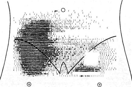
| Fig. 99. Introducing a laparoscope into the abdomen. |
Technique. The patient is placed on the operating table. The site of the puncture is treated by the common technique (usually in the median line, 3-4 cm below the navel, or in the anterolateral parts of the mesogastric region, anteriorly of the abdominal rectus muscles; Fig. 99). In order to ensure higher safety and better conditions for examination of the abdominal

Special Part
Chapter 7. Digestive System


 organs, pneumoperitoneum is first placed (oxygen, carbon dioxide or nitrous oxide are injected into the abdomen through a blunt-pointed needle). In cases with ascites, the fluid is removed. After infiltrative anaesthesia with a 0.25 per cent procaine solution, a 1-cm cut is made by a scalpel at a needed site and a trocar of the laparoscope is passed into the cut. The optical part of the laparoscope is then passed inside the trocar. The abdominal organs are inspected according to a predetermined plan. Depending on the object of examination (stomach, liver, gall bladder, etc.) the patient assumes the appropriate position on the operating table. After laparoscopy is over, air (gas) is released from the abdominal cavity, the trocar is removed, and the wound is sutured. The patient should remain in bed for three days. (The air remaining in the abdomen is absorbed within several days).
Anterosuperior and inferior surfaces of the liver can be examined during laparoscopy. The size of the liver, its colour, the character of the surface, the condition of the liver edge, and its consistency, and also a considerable part of the gall bladder are determined by laparoscopy. Laparoscopy should be done strictly for special indications. It is used in cases where other techniques fail to diagnose the disease. Laparoscopy is especially valuable in diagnosis of focal affections of the liver (tumour, cyst), in establishing the cause of ascites of unknown aetiology (cirrhosis, cancer of the liver, tuberculous peritonitis, etc.), in establishing the character of jaundice (differentiation of obstructive jaundice from hepatic one, and location of possible site and character of obstruction), in cases suspected for cancer of the gall bladder, and in splenomegaly of dubious origin.
Laparoscopy is contraindicated in severe diseases of the cardiovascular system, of the lungs, in haemorrhagic diathesis, grave forms of anaemia, and some other conditions.