Главная страница Случайная страница
КАТЕГОРИИ:
АвтомобилиАстрономияБиологияГеографияДом и садДругие языкиДругоеИнформатикаИсторияКультураЛитератураЛогикаМатематикаМедицинаМеталлургияМеханикаОбразованиеОхрана трудаПедагогикаПолитикаПравоПсихологияРелигияРиторикаСоциологияСпортСтроительствоТехнологияТуризмФизикаФилософияФинансыХимияЧерчениеЭкологияЭкономикаЭлектроника
Methods of Examination 1 страница
|
|
Inquiry
Complaints. Patients with diseases of the kidneys most commonly complain of pain in the lumbar region, disordered urination, oedema, headache, and dizziness. They may also complain of deranged vision, pain in the heart, dyspnoea, absence of appetite, nausea, vomiting, and elevated body temperature. But diseases of the kindeys may also proceed without any symptoms of renal or general clinical insufficiency.
If the patient complains of pain, its location should first of all be determined. Pain of renal origin often localizes in the lumbar region. If the ureters are affected, the pain is felt by their course. If the bladder is involved, pain is suprapubical. Radiation of pain into the perineal region is characteristic of an attack of nephrolithiasis.
The character of pain should then be determined. It is necessary to remember that the renal tissue is devoid of pain receptors. The pain is felt when the capsule or the pelvis is distended. Dull and boring pain in the lumbar region occurs in acute glomerulonephritis, abscess of the perirenal cellular tissue, in heart decompensation (" congestive kidney"), in chronic pyelonephritis (usually unilateral) and less frequently in chronic glomerulonephritis. Pain arises due to distension of the renal capsule because of the inflammatory or congestive swelling of the renal tissue. Sharp and suddenly developing pain on one side of the loin can be due to the renal infarction. The pain persists for several hours or days and then subsides gradually. The pain is rather severe in acute pyelonephritis: inflammatory oedema of the ureter interferes with the normal urine outflow from the pelvis and thus causes its distension. The pain is usually permanent. Some patients complain of attacks of severe piercing pain in the lumbar region or by the course of the ureter. The pain increases periodically and then subsides, i.e. has the character of renal colic. Obstruction of the ureter by a calculus or its bending (movable kidney) is the most common cause of this pain, which is usually attended by spasmodic contraction of the ureter, retention of the urine in the pelvis, and hence its distension. The spasmodic contractions and distension of the pelvis account for the pain. Pain in renal colic is usually unilateral. It radiates into the corresponding hypochondrium and most frequently by the course of the ureter to the
bladder and to the urethra. This radiation of pain is explained by the presence of nerve fibres (carrying the impulses from kidneys, ureters, sex organs and the corresponding skin zones) in the immediate vicinity of the relevant segments of the spinal cord (DX-DXII and LrLn). This facilitates propagation of the excitation. Patients with renal colic (like those with colic of other aetiology) are restless; they toss in bed. Patients with severe pain of other aetiology would usually lie quiet in their beds (movements may intensify the pain).
The conditions promoting pain should be established. For example, pain in nephrolithiasis can be provoked by taking much liquid, jolting motion, or the like; pain is provoked by urination in cystitis. Difficult and painful urination is observed in stranguria. Patients with urethritis feel a burning pain in the urethra during or after urination.
It is necessary also to establish the agent that lessens or removes the pain. For example, atropine sulphate, hot water-bottle or warm bath help in renal colic. Since these remedies only help in spasmodic pain by removing spasms of the smooth muscles, their efficacy in renal colic confirms the leading role of the ureter contraction in the pathogenesis of this pain. Pain of the renal colic-type in patients with movable kidney may lessen with changing posture: urine outflow improves with displacement of the kidney. Pain slightly lessens in patients with acute paranephritis if a bag with ice is placed on the lumbar region and if the patient is given amidopyrine or other analgesics.
Many renal diseases are attended by derc^l§edurmatipn: changes in the daily volume of excreted urine and in the circadian rhythm of urination.
Secretion of urine during a certain period of time is called tfijtresis. Diuresis can be positive (the amount of urine excreted exceeds the volume of liquid taken) or negative (the reverse ratio). Negative diuresis is observed in cases of liquid retention in the body or its excess excretion through the skin, by the lungs (e.g. in dry and hot weather). Positive diuresis occurs in resolution of oedema, after administration of diuretics, and in some other cases. Deranged excretion of urine is called^suria.
Increased amount of excreted urine (over 21 a day) is called golmria. It can be of renal and extrarenal aetiology. Polyuria is observed in persons who take much liquid, during resolution of oedema (cardiac or renal), and after taking diuretics. Long-standing polyuria with a high relative density of urine is characteristic of diabetes mellitus. In this case polyuria arises due to a deranged resorption of water in renal tubules because of increased osmotic pressure of the urine rich in glucose. Polyuria occurs in diabetes incipidus because of insufficient supply of antidiuretic hormone secreted into blood by the posterior pituitary. Polyuria also occurs in the absence of sensitivity of the tubules to the ADH, in affected interstice of the renal
Special Part
Chapter 8. Urinary System
 medulla of various nature, in hypokaliaemia, and hypo- and hyper-calcaemia.
Persistent polyuria with low specific gravity of urine (hyposthenuria) is usually a symptom of a severe renal disease, e.g. chronic nephritis, chronic pyelonephritis, renal arteriolosclerosis, etc. Polyuria in such cases indicates the presence of a neglected disease with renal insufficiency and decreased reabsorption in renal tubules.
Decreased amount of excreted urine (less than 500 ml a d ay) is called oliguria. It can be not connected directly with renal affections (extrarenal oliguria). For example, it can be due to limited intake of liquid, during staying in a hot and dry room, in excessive sweating, intense vomiting, profuse diarrhoea, and during decompensation in cardiac patients. But in certain cases oliguria is the result of diseases of the kidneys and the urinary ducts (renal oliguria), such as acute nephritis, acute dystrophy of the kidneys in poisoning with corrosive sublimate, etc.
A complete absence of urine secretion and excretion is called anuria. Anuria persisting for several days threatens with possible development of uraemia and fatal outcome. Anuria may be caused by the deranged secretion of urine by the kidneys (secretory anuria) which occurs in severe form of acute nephritis, nephronecrosis (poisoning with sublimate or other nephrotoxic substances), transfusion of incompatible blood, and also some general diseases and conditions such as severe heart failure, shock, or profuse blood loss.
In certain cases the secretion of urine is normal but its excretion is obstructed mechanically (obstruction of the ureters or the urethra by a calculus, inflammatory oedema of the mucosa, proliferation of a malignant tumour). This is called excretory anuria. It is usually attended by strong pain in the loin and the ureters due to distension of the renal pelves and the ureters. Exctretory anuria is often attended by renal colic.
Renal (secretory) anuria can be of reflex origin, e.g. in severe pain (contusion, fractures of the extremities, etc). Anuria should be differentiated from ischuria, when the urine is retained in the bladder and the patient is unable to evacuate it. This occurs in compression or other affection of the spinal cord, and in loss of consciousness.
Pollakiuria (frequent micturition) is observed in certain cases. A healthy person urinates from 4 to 7 times a day. The amount of excreted urine during one micturition is from 200 to 300 ml (1000-2000 ml a day). But frequency of micturition may vary within wider range under certain conditions: it may decrease in limited intake of liquid, after eating much salted food, in excessive sweating, in fever, and the like, or the frequency may increase (polyuria) if the person takes much liquid, in getting cold, and the like circumstances. Frequent desire to urinate with excretion of
meagre quantity of urine is the sign of cystitis. A healthy person urinates 4-7 times during the day time; a desire to urinate during night sleep does not arise more than once. In the presence of pollakiuria the patient feels the desire to urinate during both day and night. In the presence of chronic renal insufficiency and if the kidneys are unable to control the amount and concentration of excreted urine in accordance with the amount of liquid taken, physical exertion, the ambient temperature, or other factors important for the liquid balance in the body, the patient urinates at about equal intervals with evacuation of about equal portions of urine. This condition is called (stma.
Under certain pathological conditions, the frequency of urination is normal during the day time but increases during night. The amount of urine excreted during night often exceeds the amount of daily urine (nyc-turia). Nocturnal enuresis (nycturia) and oliguria during day time occur in cardiac decompensation and are explained by a better renal function at night, i.e. at rest (cardiac nycturia). Nycturia may concur with polyuria in renal dysfunction, at the final stage of chronic glomerulonephritis, chronic pyelitis, vascular nephrosclerosis, and other chronic renal diseases (renal nycturia). In the presence of isuria and nycturia of renal origin, which arise due to the loss by the kidneys of their concentrating ability, the specific gravity of the urine is monotonous. The condition is known as isosthenuria. The specific gravity of urine is usually decreased (hyposthenuria). The specific gravity of urine varies from 1.009 to 1.011, i.e. approaches the specific gravity of primary urine (plasma ultrafiltrate) in patients with pronounced nephrosclerosis, which is the final stage of many chronic renal diseases.
Some diseases of the bladder and the urethra are attended by difficult and painful urination. The patient would complain of change in the colour of the urine, its cloudiness, and traces of blood.
Oedema is observed in acute and chronic diffuse glomerulonephritis, nephrotic syndrome, amyloidosis, and acute renal excretory dysfunction (anuria). It is important to ask the patient about the site that was the first to be attacked by oedema, the sequence of oedema spreading, and the rate of intensification of this phenomenon (see " Renal Oedema").
Headache, dizziness, and heart pain may result from kidney affections. These symptoms occur in those renal diseases which are attended by considerable increase in the arterial pressure, e.g. in acute and chronic glomerulonephritis or vascular nephrosclerosis. A pronounced and persistent increase in the arterial pressure can be among the causes of deranged vision (neuroretinitis).
Patients with diseases of the kidneys can complain of weakness, indisposition, impaired memory and work capacity and deranged sleep. Vi-
Special Part
Chapter 8. Urinary System


 sion may be deranged along with skin itching and unpleasant breath. Dyspeptic disorders sometimes join in: loss of appetite, dryness and unpleasant taste in the mouth, nausea, vomiting, and diarrhoea. All these phenomena are associated with retention in the body of protein decomposition products due to renal insufficiency (see " Renal Insufficiency") which develops at the final stage of many chronic renal diseases, and sometimes in acute diseases attended by retention of urine during several days.
Fever is the common symptom of infectious inflammatory affections of the kidneys, the urinary ducts and perirenal cellular tissue.
History of the present disease. When questioning the patient, it is necessary to establish the connection of the present disease with previous infections (tonsillitis, scarlet fever, otitis, acute respiratory diseases). This sequence is especially characteristic of acute glomerulonephritis. But it is sometimes difficult to establish the time of onset of the disease because some chronic affections of the kidneys and the urinary ducts can for a long time be latent. Moreover, when questioning the patient, it is necessary to find out if he had deranged hearing or vision in his childhood that might be suggestive of congenital renal pathology.
Special attention should be given to the presence in the patient's past history of diseases of the kidneys and the urinary ducts (acute nephritis, pyelitis, cystitis) or symptoms that might suggest them (dysuria, haematuria, oedema, arterial hypertension, attacks of pain in the abdomen or loin resembling renal colics), since these symptoms can be connected with the present renal pathology. In certain cases the cause and the time of onset of grave kidney affections (necronephrosis) can be established by revealing industrial or domestic poisoning, intentional (or by mistake) taking of some poisons (corrosive sublimate, preparations of bismuth, phosphorus, silver, large doses of sulpha preparations, or of some antibiotics, e.g. aminoglycosides, expired tetracyclines, phosphorus compounds), transfusion of incompatible blood, etc. Amidopyrin, phenacetin, barbiturates, camphor, and some other medicines can cause allergic changes in the kidneys.
The patient must be asked about the character of the disease course: it may be gradual (arteriolosclerosis, chronic diffuse glomerulonephritis, amyloidosis of the kidneys), or with periodical exacerbations (chronic pyelonephritis, chronic diffuse glomerulonephritis). It is necessary to establish the cause of exacerbations, their frequency, clinical signs, the character of therapy given and its efficacy, the causes inducing the patient to seek medical help.
Anamnesis. Special attention should be given to the factors that might provoke the present disease or have effect on its further course. For example, a common factor promoting development of acute and chronic
nephritis and pyelonephritis is chilling and cooling (poor housing or working conditions, drafts, work in the open, acute cooling of the body before the disease). Spreading of genital infection onto the urinary system can be the cause of pyelonephritis. It is necessary to establish the presence or absence in the past of tuberculosis of the lungs or other organs. This helps establish the tuberculous nature of the present disease of the kidneys.
It is necessary to establish if the patient has some other diseases that might cause affections of the kidneys (collagenosis, diabetes mellitus, certain diseases of the blood, etc.). Various chronic purulent diseases (osteomyelitis, bronchiectasis) can be the cause of amyloidosis of the kidneys. Occupations associated with walking, riding, weight lifting, etc., can have their effect on the course of nephrolithiasis and provoke attacks of renal colic. Some abnormalities of the kidneys, nephrolithiasis, amyloidosis, etc., can be inherited. It is also necessary to record thoroughly the information on past operations on the kidneys or the urinary ducts.
When examining women, it is important to remember that pregnancy can aggravate some chronic diseases of the kidneys and be the cause of the so-called nephropathy of pregnancy (toxaemia of late pregnancy).
Physical Examination
INSPECTION
Inspection of the patient should give the physician the idea of the gravity of the patient's condition. Very grave condition with loss of consciousness may be due to severe affections of the kidneys attended by renal insufficiency and uraemic coma; the condition may be satisfactory or of moderate gravity (in milder cases). It is necessary to pay attention to the patient's posture in bed: active (at initial stages of many diseases of the kidneys), passive (in uraemic coma), or forced (in paranephritis; the patient may lie on his side with the leg flexed, bringing the knee to the abdomen on the affected side). In the presence of renal colic the patient is restless, tosses in bed, groans or even cries from pain. Convulsions are observed in the presence of uraemic coma, renal eclampsia, and nephropathy of pregnancy (toxaemia of late pregnancy with involvement of the kidneys).
Oedema is characteristic of acute and chronic glomerulonephritis, nephrotic syndrome, and amyloidosis of the kidneys. The appearance of the patient with oedema of the renal origin is quite specific (Fig. 101). The face is pallid, swollen, with oedematous eyelids and narrowed eye-slits (facies nephritica). In patients with more pronounced signs of pathology, oedema affects the upper and lower extremities and the trunk (anasarca).
The colour of the patient's skin is also important. Oedematous skin in
Special Part
Chapter 8. Urinary System


chronic nephritis is pallid due to the spasm of skin arterioles, and anaemia which attends this disease. The skin is wax-pallid in amyloidosis and lipoid nephrosis. It should be remembered that in cardiac oedema (as distinct from renal oedema) the skin is more or less cyanotic.
When inspecting a patient with chronic nephritis, it is possible to observe scratches on the skin and coated dry tongue; an unpleasant odour of ammonia can be felt from the mouth and skin of the patient (factor uremicus). All these signs characterize chronic renal insufficiency (uraemia).
Inspection of the abdomen and the loin does not usually reveal any noticeable changes. But in the presence of paranephritis, it is possible to notice swelling on the affected side of the loin. In rare cases, an especially large tumour of the kidney may be manifested by protrusion of the abdominal wall. Distended bladder can be protruded over the pubic bone in thin persons. The distension can be due to overfilling of the bladder, for example, due to retention of urine in adenoma or cancer of the prostate.
PALPATION
The posterior location of the kidneys, and also the absence of anterior approach to them due to the interference of the costal arch, makes palpation of the kidneys difficult. Relaxation of the prelum and pronounced
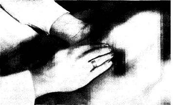
Fig. 102. Palpation of the right kidney of the lying patient. -I
cachexia can be attended by certain ptosis of the kidneys and make them accessible to palpation even in healthy subjects. But the results of palpation can only be reliable in considerable enlargement of the kidneys (at least 1.5—2 times, e.g. due to formation of a cyst or a tumour), or their displacement by a tumour, or in cases with a floating kidney. Bilateral enlargement of the kidneys is observed in polycystosis.
It is necessary to remember that the kidneys can move about in the range of 2-3 cm in the proximal and distal directions when the subject changes his position from horizontal to vertical, and also during respiratory movements of the diaphragm. Passive movements of the kidneys transmitted from the diaphragm during inspiration and expiration should be taken into consideration during palpation: the Obraztsov-Strazhesko palpation method should be used. The patient should be palpated in the lying or standing position. When the patient is in the horizontal position, his kidneys sfre better palpated because the strain of the prelum is absent. But the movable kidney can be palpated in the standing patient because it hangs by gravity and is displaced downward by the pressure of the low diaphragm.
During palpation of the patient in the lying position (Fig. 102), his legs should be stretched and the head placed on a low pillow; the prelum is relaxed and the arms are freely placed on the chest. The physician should assume his position by the right side of the patient with his left hand under the patient's loin, slightly below the 12th rib so that the finger tips be near the spinal column. During palpation of the left kidney, the physician's
Special Part
Chapter 8. Urinary System
hand should be moved further, beyond the vertebral column, to reach the left part of the lumbar region. The right hand should be placed on the abdomen, slightly below the corresponding costal arch, perpendicularly to it and somewhat outwardly of the rectus abdominis muscles. The patient is asked to relax the abdominal muscles as much as possible and breathe deeply and regularly. The physician's right hand should press deeper with each expiration to reach the posterior abdominal wall, while the left hand presses the lumbar region to meet the fingers of the right hand. When the examining hands are as close to each other as possible, the patient should be asked to breathe deeply by " the abdomen" without straining the prelum. The lower pole of the kidney (if it is slightly descended or enlarged) descends still further to reach the fingers of the right hand. As the physician feels the passing kidney, he presses it slightly toward the posterior abdominal wall and makes his fingers slide over the anterior surface of the kidney bypassing its lower pole. If ptosis of the kidney is considerable, both poles and the entire anterior surface of the kidney can be palpated. The physician should assess the shape, size, surface (smooth or tuberous), tenderness, mobility, and consistency of the kidneys. Bimanual palpation of the kidney can also be done with the patient lying on his side.
In contrast to other organs, an enlarged or ptosed kidney can be examined by ballottement (Guyon's sign): the right hand feels the kidney while the fingers of the left hand strike rapidly the lumbar region in the angle between the costal arch and the longissimus thoracic muscles: the fingers of the right hand feel vibration of the kidney. In deranged urine outflow through the ureter and in pronounced distension of the renal pelvis by the accumulated urine or pus, liquid fluctuation can be felt during palpation of the kidney.
If the physician palpates some formation where he expects to find a kidney, he must check reliably if this is actually a kidney because it is easy to mistake for the kidney an overfilled and firm part of the large intestine, tumor of perirenal cellular tissue (lipoma, fibroma), an enlarged right lobe of the liver, the gall bladder (during palpation of the right kidney), or an enlarged or displaced spleen (during palpation of the left kidney). The kidney is a bean-shaped body with a smooth surface, slipping upwards from under the palpating fingers and returning to normal position, tossed up by ballottment and giving tympany during percussion over the kidney (by overlying intestinal loops). Protein and erythrocytes appear in the urine after palpation. But all these signs are of only relative importance. For example, if a malignant tumour develops, the kidney may lose its mobility due to proliferation of the surrounding tissues; its surface becomes irregular and the consistency more firm; if the tumour is large, the kidney moves apart the intestinal loops and percussion gives dullness. But the
kidney can nevertheless be identified by the mentioned signs by differentiating it from the neighbouring organs and other formations.
Palpation of the kidneys in the standing patient was proposed by S. Botkin. During palpation the patient stands facing the physician who sits on a chair. The prelum muscles should be relaxed and the trunk slightly inclined forward.
Palpation can be used to diagnose ptosis of the kidneys. Three degrees of nephroptosis can be distinguished: the lower pole of the kidney can be palpated in cases with ptosis of the first degree; the entire kidney can be palpated in the second degree; and the kidney freely moves about in all directions to pass beyond the vertebral column, to the side of the other kidney, and to sink downwards to a considerable distance, in the third-degree ptosis.
Palpation is also used to examine the bladder. If it contains much urine, especially in persons with thin abdominal wall, the urinary bladder can be palpated over the pubic bone as an elastic fluctuating formation. If the bladder is markedly distended, its superior border reaches the umbilicus.
Tenderness in palpation of the ureter along its course and sensitive loin over the kidneys (sensitive to pressure exerted in the angle between the 12th rib and the longissimus thoracic muscles) is of certain diagnostic importance. The area overlying the ureter extends on the anterior abdominal wall between the superior ureter point (at the edge of the rectus abdominis muscle at the level of the umbilicus) and the inferior point (at the intersection of the bi-iliac line and the vertical line passing the pubic tubercle).
PERCUSSION
It is impossible to percuss the kidneys in a healthy subject because they are covered anteriorly by the intestinal loops which give tympany. Dullness can only be determined in the presence of very marked enlargement of the kidneys.
A much more informative method for examination of the kidneys is tapping. The physician places his left hand on the patient's loin and using his right hand (palm edge or fingers) taps with a moderate force on the right hand overlying the kidney region on the loin (Fig. 103). If the patient feels pain, the symptom is positive (Pasternatsky's symptom). This symptom is also positive in nephrolithiasis, paranephritis, inflammation of the pelvis, and also in myositis and radiculitis. This decreases the diagnostic value of Pasternatsky's symptom.
A full urinary bladder gives a dull sound on percussion of the suprapubic region. The percussion is carried out from the umbilicus downward; along the median line; the pleximeter-finger is placed parallel to the pubic bone.
Special Part
Chapter 8. Urinary System
|
i!
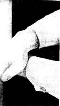
Fig. 103. Determining Pasternatsky's symptom.
Instrumental and Laboratory Methods
URINALYSIS
The study of urine is important for establishing a diagnosis of and concluding on the course of the pathology. Various pathological processes occurring in the kidneys and the urinary tracts have their effect on the properties of urine. Pathological metabolites may be released into the blood in various diseases. Excreted by the kidneys, these metabolites are also found in the urine and their determination is therefore important diagnostically. Urine samples taken after night sleep are usually studied. The analysis begins with the study of its physical properties.
The normal daily amount of urine (daily diuresis) excreted by an adult varies from 1000 to 2000 ml, the ratio of the urine evacuated during the day to the nocturnal diuresis being 3: 1 or 4: 1. The daily amount of urine below 500 ml and over 2000 ml can be considered pathological under certain conditions.
The colour of normal urine depends on its concentration and varies from straw-yellow to the colour of amber. Concentration of urochromes, urobilinoids, uroerythrin and of some other substances accounts for the colour of urine. The most marked changes in the urine colour depend on the presence of greenish-brown bilirubin, large quantity of erythrocytes (appearance of meat wastes), reddish-brown urobilin, and medicines (acetylsalicylic acid and amidopyrine give pink colour to the urine, methylene blue colours it blue, and rhubard greenish-yellow). Normal
urine is clear. Cloudiness may be due to salts, cell elements, mucus, fats, and bacteria.
The smell of urine is specific and not pungent. When decomposed by bacteria in- or outside the bladder, urine smells of ammonia. In the presence of ketone bodies (in grave forms of diabetes mellitus), urine smells " fruity" (the odour of decomposing apples).
The specific gravity of the urine varies from 1.001 to 1.040. It is measured by an urometer (hydrometer) with the scale reading from 1.000 to 1.050. Determination of the specific gravity of the urine is of great clinical importance because it gives information on the concentration of substances dissolved in it (urea, uric acid, salts) and characterizes the concentrating and diluting capacity of the kidneys. It should be remembered that specific gravity depends not only on the amount of particles dissolved but mainly on their molecular weight. High-molecular substances (e.g. proteins) account for increased specific gravity of the urine without influencing substantially the osmotic concentration of the urine. The osmotic concentration of the urine depends mainly on the presence of electrolytes and urea. Osmotic concentration is expressed in mosm/1. The maximum osmotic concentration of urine in a healthy person is 910 mosm/1 (maximum sp. gravity, 1.025-1.028). The specific gravity of the urine may exceed 1.030-1.040 in the presence of high quantity of glucose (glucosuria), because the concentration of 10 g/1 increases gravity of the urine by 0.004.